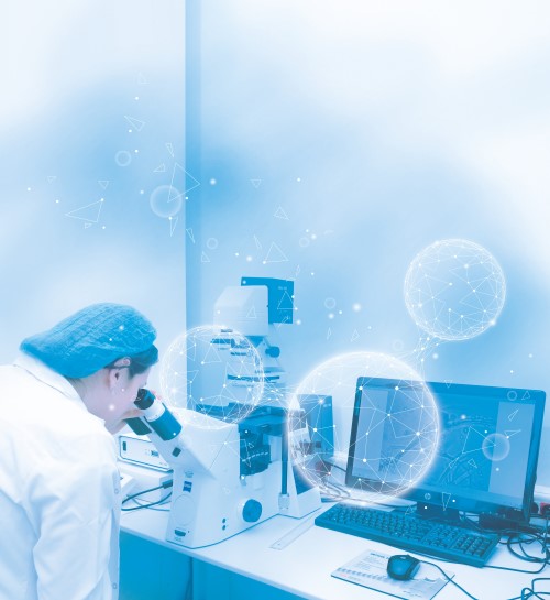
In our Imaging Laboratory, the following studies are carried out using the microscopes that provide the analysis, processing and three-dimensional configuration of images with comprehensive methods:

Phase Contrast Microscope is used to perform the following Imagings:
• Negative Staining Display
• Tissue, Section, Print, Smear
• Tissue Section Screening
• Filamentlike Dust Count
• Viability - Apoptosis (Stained with Fluorescent Dye)
• Histologic Inspection
• Mold Identification.
Stereo microscope is used to perform the following:
• Macroscopic
• Microplastic
• Foreign Matter Identification.
Optical microscope is used to perform the following:
• Gram Stain Imaging
• Capsule Staining Imaging
• Spore Staining Display
• Cell Count
• Parasite Search is in progress.
Our highly equipped devices and technical materials we use are calibrated periodically by accredited calibration institutions in accordance with the legislation.
In addition, we periodically participate in national and international qualification and comparison tests with a goal of 100% success in order to guarantee the reliability of our analysis methods.
